
Key take‑aways
- Blockage raises ductal pressure, sparking chronic inflammation and fibrosis.
- Exocrine output drops, leading to malabsorption and weight loss.
- Endocrine cells suffer, increasing the risk of type3c diabetes.
- Early imaging (MRCP/ERCP) and timely drainage can slow or reverse damage.
- Long‑term management hinges on enzyme replacement, glucose monitoring, and lifestyle adjustments.
What is pancreatic duct obstruction?
Pancreatic duct obstruction is a blockage of the main conduit that carries digestive enzymes from the pancreas into the duodenum. The obstruction can arise from gallstones, scar tissue, tumors, or strictures, and it forces pancreatic secretions to back‑up, elevating intraductal pressure.
When pressure builds, the pancreas essentially tries to push against a closed door. The immediate result is a cascade of cellular stress that, over months or years, reshapes how the organ works.
How the blockage hijacks exocrine function
Exocrine pancreatic function is a type of digestive activity that releases enzymes like amylase, lipase, and proteases into the small intestine. Its main goal is to break down carbs, fats, and proteins so the body can absorb nutrients.
With a blocked duct, those enzymes can’t reach the gut. Instead, they sit inside the gland, become activated prematurely, and start digesting pancreatic tissue itself. Over time, the gland loses its enzyme‑producing cells, a condition known as exocrine pancreatic insufficiency (EPI). Patients with EPI commonly report greasy stools, bloating, and unintended weight loss.
Impact on endocrine health - the hidden diabetes risk
Endocrine pancreatic function refers to the hormone‑producing cells (islets of Langerhans) that regulate blood sugar by releasing insulin and glucagon.
The same pressure that damages exocrine cells also compresses the delicate islets. Chronic inflammation and fibrosis create a hostile environment, gradually killing beta‑cells. This specific form of diabetes, sometimes called type3c diabetes, differs from type1 and type2 because it stems from pancreatic disease rather than autoimmunity or insulin resistance.
From inflammation to chronic pancreatitis
Chronic pancreatitis is a long‑standing inflammation of the pancreas that leads to irreversible tissue damage.
The ductal blockage acts as a relentless irritant. Repeated bouts of acute inflammation evolve into chronic pancreatitis, marked by persistent pain, calcifications, and loss of both exocrine and endocrine tissue. Studies from major UK gastroenterology centres show that up to 60% of patients with longstanding ductal obstruction develop chronic pancreatitis within a decade.
Fibrosis - the scar tissue that locks the pancreas down
Pancreatic fibrosis is the deposition of collagen and other extracellular matrix proteins that replace healthy pancreatic cells.
Fibrosis is the body’s wound‑healing response gone awry. In the pancreas, it stiffens the gland, further impedes ductal flow, and seals off the remaining functional tissue. Imaging studies (MRI, CT) often reveal a “sausage‑shaped” pancreas with irregular borders in patients with longstanding obstruction.
Clinical fallout: malabsorption and weight loss
Malabsorption syndrome occurs when the small intestine cannot absorb nutrients efficiently, leading to deficiencies in vitamins A, D, E, K and essential fatty acids.
When the exocrine pancreas fails, fats remain undigested, pulling bile acids into the lumen and causing steatorrhea (fatty stools). Over months, patients lose up to 15% of body weight, develop anemia, and may suffer bone demineralisation due to vitamin D loss.
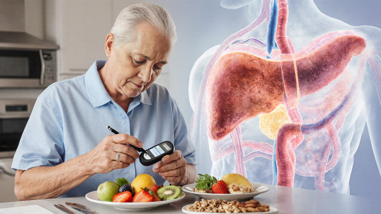
Diabetes type3c - a silent consequence
Diabetes mellitus (type3c) is a form of diabetes that arises secondary to pancreatic disease, such as chronic pancreatitis or pancreatic cancer.
Because the pancreas can no longer produce enough insulin, blood sugars climb. Unlike classic type2 diabetes, patients often have a blunted glucagon response, making them prone to both hyper‑ and hypoglycaemia. Regular HbA1c monitoring and tailored insulin regimens become essential.
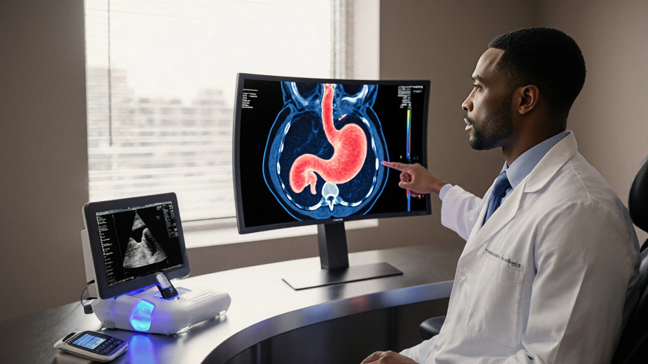
Diagnosing a blocked duct - imaging and labs
Modern imaging offers a clear picture of ductal anatomy. Magnetic resonance cholangiopancreatography (MRCP) provides non‑invasive 3‑D maps, while endoscopic retrograde cholangiopancreatography (ERCP) doubles as a therapeutic tool. Lab work typically shows elevated serum amylase, lipase, and low fecal elastase (a marker of exocrine insufficiency).
Treatment options - draining the pressure
Two main strategies aim to restore flow:
| Attribute | Endoscopic Stenting | Surgical Drainage |
|---|---|---|
| Success rate (short‑term) | ≈85% | ≈95% |
| Invasiveness | Minimally invasive (via duodenum) | Requires laparotomy or laparoscopy |
| Complication risk | Post‑ERCP pancreatitis (3‑5%) | Infection, bleeding (≈7%) |
| Recovery time | 1‑2days hospital stay | 5‑7days hospital stay |
| Long‑term patency | Stent occlusion possible (12‑18months) | Usually durable; may need revision |
Endoscopic stenting involves placing a tiny tube across the narrowed segment during ERCP, allowing secretions to bypass the blockage.
Surgical drainage (e.g., pancreaticojejunostomy) creates a permanent connection between the pancreas and the jejunum, fully diverting flow.
Choosing between them depends on patient age, comorbidities, and the anatomical location of the obstruction. Younger patients with localized strictures often fare better with surgery, while high‑risk patients benefit from endoscopic approaches.
Long‑term management beyond the procedure
Even after successful drainage, the pancreas may never fully recover. Ongoing care includes:
- Pancreatic enzyme replacement therapy (PERT) - doses tailored to weight and stool fat content.
- Regular monitoring of blood glucose and HbA1c to catch type3c diabetes early.
- Vitamin supplementation (A, D, E, K) to offset malabsorption.
- Low‑fat, high‑protein diet to maximise nutrient uptake.
- Annual imaging to watch for progressive fibrosis or neoplastic changes.
Patients who maintain enzyme therapy and glycaemic control often report improved quality of life and reduced hospital admissions.
Prevention - spotting the warning signs early
Because many blockages stem from gallstones or alcohol‑related pancreatic injury, lifestyle tweaks can lower risk:
- Limit alcohol to ≤14units/week; abstain if you already have chronic pancreatitis.
- Adopt a diet rich in omega‑3 fatty acids to reduce biliary sludge formation.
- Stay vigilant for recurrent upper‑abdominal pain, especially after fatty meals.
- Seek medical attention if you notice persistent oily stools or unexplained weight loss.
Early imaging and enzyme testing can catch a nascent blockage before fibrosis sets in.
Future directions - where research is heading
Emerging therapies aim to halt fibrosis. Trials of monoclonal antibodies targeting transforming growth factor‑beta (TGF‑β) show promise in reducing scar formation. Additionally, endoscopic laser lithotripsy is being refined to break down stubborn stones that cause obstruction without the need for open surgery.
Genetic profiling may soon identify patients predisposed to aggressive fibrosis, allowing for personalised surveillance intervals.
Frequently Asked Questions
Can a blocked pancreatic duct cause cancer?
Chronic inflammation from a blocked duct raises the risk of pancreatic ductal adenocarcinoma. While the absolute risk remains low, patients with long‑standing obstruction should undergo regular imaging to catch any suspicious lesions early.
What symptoms signal a pancreatic duct blockage?
Typical signs include persistent upper‑abdominal pain that radiates to the back, greasy or frothy stools, unexplained weight loss, and occasional episodes of nausea after fatty meals. Lab tests often reveal low fecal elastase alongside elevated serum amylase.
Is surgery always required for a blocked duct?
No. Endoscopic stenting can relieve pressure in many cases, especially when the blockage is due to a short stricture or removable stone. Surgery is reserved for failures of endoscopic therapy or when anatomy precludes a safe stent placement.
How effective is pancreatic enzyme replacement therapy?
When dosed correctly, PERT normalises stool fat content in up to 80% of patients with exocrine insufficiency. Dose adjustments based on symptom response and periodic fecal fat analysis optimise outcomes.
Can lifestyle changes reverse the damage?
Lifestyle tweaks can halt further injury but rarely reverse established fibrosis. However, they improve overall pancreatic health, reduce pain episodes, and enhance the effectiveness of medical therapies.


Fatima Sami
The article contains a few grammatical oversights. For instance, the phrase “exocrine output drops” should be preceded by “the” to read “the exocrine output drops”. Additionally, “increase the risk of type3c diabetes” would read more clearly as “increases the risk of type 3c diabetes”. Consistency in hyphenation-such as “type‑3c” versus “type3c”-should be standardised throughout.