
Ever wondered what's actually happening inside your eye during those long appointments where you have to stare at a blinking light? If you've ever had a "scan" of your retina, you've likely encountered Ophthalmic Imaging. It sounds technical, but it's basically the difference between seeing a blurry photo of a forest and having a 3D map of every single leaf and root. These tools don't just look at the surface; they let doctors peer through layers of tissue to find problems long before you notice a change in your vision.
The Big Three: What are these tests?
When a doctor wants to check your retinal health, they usually rely on a combination of tools. Think of it like a crime scene investigation: one tool captures the wide shot, another looks for blood flow, and a third provides a microscopic cross-section. The three main players are Fundus Photography, Fluorescein Angiography, and Optical Coherence Tomography (OCT).
Fundus Photography is a specialized camera technique that takes a high-resolution digital image of the back of the eye. This includes the retina, the optic disc, and the macula. It's essentially a high-def photo of your eye's "wallpaper." While it's great for documenting the overall state of the eye, it only shows the surface. If there's a leak happening deep inside a layer of tissue, a photo won't always catch it.
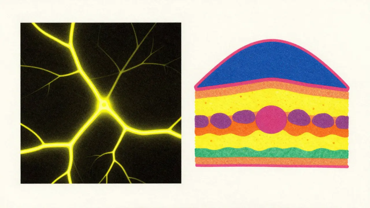
Fluorescein Angiography (FA) is a diagnostic procedure where a yellow dye is injected into the arm to highlight the blood vessels in the retina. Developed back in the 1960s, it's the gold standard for seeing "leakage." If a vessel is leaking fluid-common in diabetic retinopathy-the dye will pool in those areas, making the problem glow under a special filter. However, it's invasive, takes 10 to 30 minutes, and carries a small risk of allergic reactions to the dye.
Optical Coherence Tomography (or OCT) is a non-invasive imaging test that uses light waves to take cross-sectional views of the retina. If Fundus Photography is a photo, OCT is an ultrasound, but with light instead of sound. It allows doctors to see the individual layers of the retina and the choroid. Since the late 2000s, Spectral-Domain OCT (SD-OCT) has become the standard, offering axial resolutions as sharp as 5-7μm.
Comparing the Imaging Methods
You might wonder why a doctor wouldn't just use one of these. The truth is, they all see different things. While OCT is incredible for anatomy (the "where" and "what"), FA is better for dynamics (the "how it's leaking").
| Feature | Fundus Photo | Fluorescein Angiography (FA) | OCT / OCTA |
|---|---|---|---|
| Invasiveness | Non-invasive | Invasive (Dye injection) | Non-invasive |
| Primary Use | Surface documentation | Blood flow & leakage | Layer-by-layer anatomy |
| Time Required | Seconds | 10-30 Minutes | Seconds to Minutes |
| Best For... | General screening | Diabetic Macular Edema | Macular holes, Fluid pockets |
The New Player: OCT Angiography (OCTA)
Around 2014, a game-changer arrived: OCT Angiography (OCTA). This is essentially a hybrid. It gives you the blood vessel mapping of an angiography but without the needles and dyes. It works by detecting the motion of red blood cells in the vessels.
OCTA is a huge win for patients who are afraid of needles or have dye allergies. It can map the superficial, middle, and deep capillary plexuses of the fovea in seconds. In some cases, like detecting neovessels in proliferative diabetic retinopathy, studies have shown OCTA can actually be superior to traditional FA. For instance, a study of 30 eyes with severe diabetic retinopathy found that OCTA was more effective at spotting neovascularization of the disc (NVDs).
However, it's not perfect. Because it relies on motion, if you blink or twitch during the scan, you get "motion artifacts"-essentially glitches in the image. Also, unlike FA, OCTA can't see if fluid is actually leaking out of a vessel; it only sees if the vessel is there and if blood is moving through it.
Diagnosing Specific Eye Conditions
Depending on what the doctor is looking for, they'll lean on different images. Let's look at how this works in real-world scenarios.
Diabetic Retinopathy
For patients with diabetes, the goal is to find microaneurysms (tiny bulges in blood vessels) and edema (swelling). While OCTA is great for finding the vessels, Fluorescein Angiography often remains the top choice for detecting low-grade leakage. In some cases of diabetic macular edema, FA has shown higher sensitivity than SD-OCT because mild leaks might not change the thickness of the retina enough for an OCT scan to flag them as a problem.
Age-Related Macular Degeneration (AMD)
In AMD, doctors look for "wet" vs "dry" forms. OCT is the hero here. It can show the exact moment fluid begins to accumulate under the retina, allowing doctors to start injections (anti-VEGF therapy) before permanent vision loss occurs. Swept-source OCT (SS-OCT) is even better for this, as it can penetrate deeper into the choroid, scanning up to 400,000 A-scans per second compared to the 85,000 of older SD-OCT machines.
Rare Conditions: Coats Disease and PIC
In rarer cases like Coats Disease, OCT reveals things that photos and FA completely miss. In about 82% of studied eyes, OCT found exudates (leaky fats/proteins) hidden in multiple retinal layers. For Punctate Inner Choroidopathy (PIC), OCTA has become essential because it can detect "non-perfusion" areas-spots where blood simply isn't flowing-which are often invisible on traditional scans.

What to Expect During Your Visit
If you're heading in for imaging, here's the reality of the workflow. Fundus photos and OCTs are quick; you'll sit at a machine, look at a target, and be done in a few minutes. They are generally unaffected by your pupil size or how clear your eye's natural lens is (like if you have a slight cataract).
If you need an FA, expect a longer process. You'll have a dye injected into your arm, and the technician will take a series of photos over 10 to 30 minutes as the dye moves through your system. It's a bit more taxing and requires a stable environment.
The biggest challenge for any of these tests? Staying still. Whether it's the high-speed scans of an SS-OCT or the motion-sensitive OCTA, the quality of your results depends on your ability to keep your eye fixed on the target. This is why children or patients with advanced disease often find these tests frustrating.
Does an OCT scan hurt?
No, OCT is completely non-invasive and non-contact. No part of the machine touches your eye; it simply uses light waves to create a map of your retina. You may feel a bright light, but there is no pain involved.
Why do I need an angiography if I already had an OCT?
OCT shows the anatomy (the structure), but angiography shows the activity. While OCT can see that fluid is present (edema), angiography tells the doctor if that fluid is actively leaking from a blood vessel in real-time. They provide different, complementary pieces of the puzzle.
What are the risks of the dye used in angiography?
Fluorescein dye can cause a mild allergic reaction in some people. In rare cases, more severe reactions can occur. This is why doctors ask about allergies beforehand. Side effects can also include a temporary yellowing of the skin or urine after the procedure.
How often do I need these scans?
This depends on your condition. For routine monitoring of chronic issues like glaucoma or stable AMD, scans might happen every 6 to 12 months. For active conditions requiring injections, you might have an OCT every few weeks to track the effectiveness of the treatment.
Can OCTA replace traditional angiography?
In many cases, yes, because it's faster and safer. However, it cannot yet fully replace FA because it doesn't show leakage patterns. Doctors will often use both if they need a complete picture of the vascular health.
Next Steps for Your Eye Health
If you've been scheduled for these tests, don't stress. Most are incredibly fast. If you're prone to movement or have trouble focusing, let your technician know so they can help stabilize your head. If you're getting an FA, remember to stay hydrated and mention any history of allergies to the nursing staff.
Once you get your results, ask your doctor to show you the images. Seeing the cross-section of your retina on an OCT scan often makes it much easier to understand why a specific treatment, like an injection or laser therapy, is necessary. If you're managing a long-term condition, keep a record of your OCT thickness measurements-this data helps track whether your disease is stabilizing or progressing over time.


